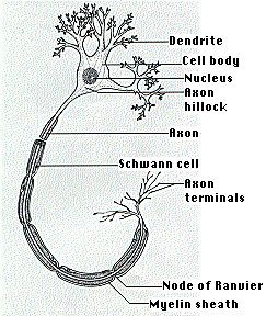
All neurons outside the central nervous system (and many within it) conduct impulses along hairlike cytoplasmic extensions, the nerve fibers or axons. (The diagram represents a motor neuron with most of its axon omitted.) The axons connecting your spinal cord to your foot can be as much as 1 m long (although only a few micrometers in diameter).
Axons grow out of the cell body, which houses the nucleus as well as other organelles such as the endoplasmic reticulum. The length of some axons is so great that it is remarkable that the cell body controls them all the way to their tip. There is a steady transport of cell components (e.g., vesicles, mitochondria) from the cell body along the entire length of the axon. This flow is driven by kinesins moving along the many microtubules in the cytoplasm within the axon. Even so, it may take 2 weeks or longer for material synthesized in the cell body to reach the axon terminals in your big toe.
In many neurons, nerve impulses are generated in short branched fibers called dendrites and also in the cell body. The impulses are then conducted along the axon, which usually branches several times close to its end.
Many axons are covered with a glistening fatty sheath, the myelin sheath. It is the greatly-expanded plasma membrane of an accessory cell, the Schwann cell. Schwann cells are spaced at regular intervals along the axon. Their plasma membrane is wrapped around and around the axon forming the myelin sheath.
Where the sheath of one Schwann cell meets that of the next, the axon is unprotected. This region, the node of Ranvier, plays an important part in the propagation of the nerve impulse.
The cell bodies of the sensory neurons leading to the spinal cord are located in clusters, the dorsal root ganglia (DRG), next to the spinal cord. Their axon extends in both directions: a peripheral axon to receptors at the periphery and a central axon passing into the spinal cord. The latter axon usually terminates at an interneuron.
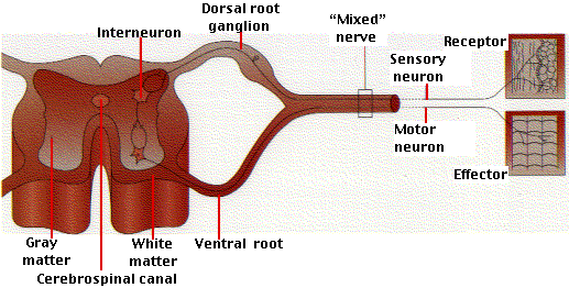
The diagram is a simplified view of the relationship between sensory and motor neurons running to and from the spinal cord.
Sensory neurons
These run from the various types of stimulus receptors, e.g.,
touch
odor
taste
sound
vision
to the central nervous system (CNS), the brain and spinal cord.
So, for example, if you stub your toe, nerves in your toe send the message to your brain through sensory neurons.
Interneurons
These are found exclusively within the spinal cord and brain. They are stimulated by signals reaching them from
sensory neurons other interneurons or both.Interneurons are also called association neurons.
It is estimated that the human brain contains 100 billion (1011) interneurons averaging 1000 synapses on each!
The term interneuron hides a great diversity of structural and functional types of cells. In fact, it is not yet possible to say how many different kinds of interneurons are present in the human brain. Certainly hundreds; perhaps many more.
Motor neurons
These transmit impulses from the central nervous system to the muscles and glands that carry out the response.
Most motor neurons are stimulated by interneurons, although some are stimulated directly by sensory neurons.
So, for example if you want to kick a ball, your brain sends a message to your leg muscles to kick it, through motor neurons.